Echocardiography in Dehradun
If you’ve read through the earlier pages on this site, you’ve probably noticed one test mentioned again and again — the echocardiogram. It comes up so often because it’s genuinely the single most useful test for looking directly at how the heart is structured and how well it’s working, without needing to go anywhere near a needle or a catheter. This page explains what an echo actually checks, how it’s done, and how to make sense of the report afterward.
Dr. Richa Sharma is an Interventional Cardiologist practicing in Dehradun, using echocardiography as a core part of evaluating almost every type of heart condition, from valve disease to heart failure to unexplained symptoms.
What Is an Echocardiogram?
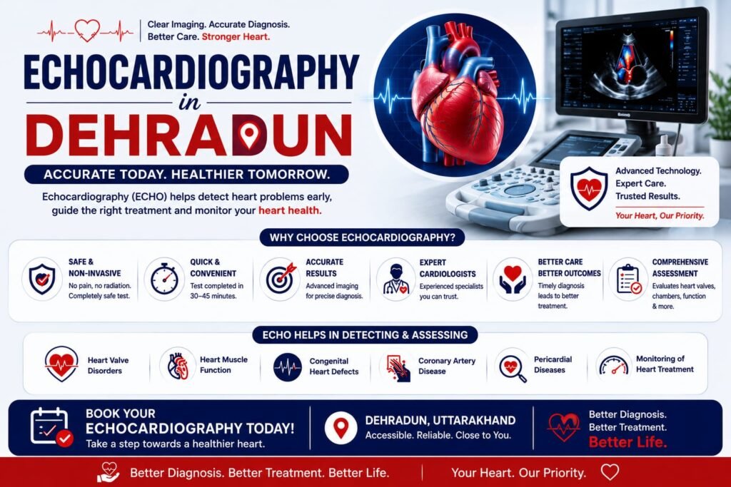
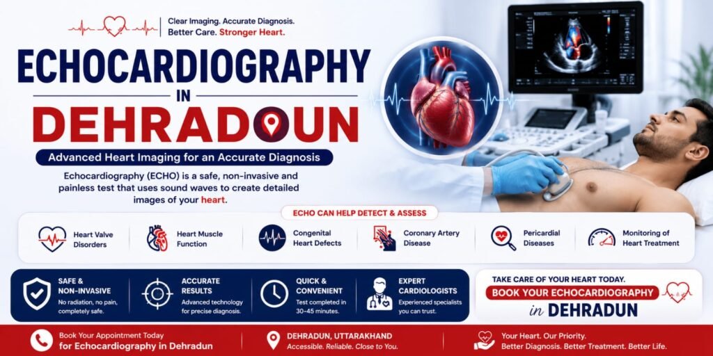
An echocardiogram, often just called an “echo,” is an ultrasound scan of the heart. It uses sound waves, not X-rays or radiation, to create real-time moving images of the heart’s chambers, valves, and the way blood flows through them. Because it shows the heart actually beating, it gives information that static tests like an ECG or a blood test simply can’t provide.
A standard echo is often referred to as a “2D echo,” since it produces two-dimensional images, though most modern machines also capture additional information like blood flow direction and speed using a technique called Doppler ultrasound, all within the same test.
What Does an Echocardiogram Check?
Pumping function (ejection fraction) — how much blood the heart pumps out with each beat, a key measure used in diagnosing and monitoring heart failure.
Heart valve function — whether any valve is narrowed (stenosis) or leaking (regurgitation), and how severely.
Chamber size and wall thickness — whether any part of the heart has enlarged or thickened, which can indicate long-standing high blood pressure, valve disease, or other conditions.
Heart muscle movement — whether all parts of the heart muscle are contracting normally, which can reveal areas damaged by a past heart attack.
Fluid around the heart — checking for any abnormal fluid buildup in the sac surrounding the heart (pericardial effusion).
Congenital abnormalities — structural heart differences present from birth, in both children and adults.
Blood clots, in some cases, particularly within the heart’s chambers.
Because it covers so much ground in a single test, an echo is often one of the very first investigations ordered once a cardiologist suspects almost any structural or functional heart problem.
When Is an Echocardiogram Recommended?
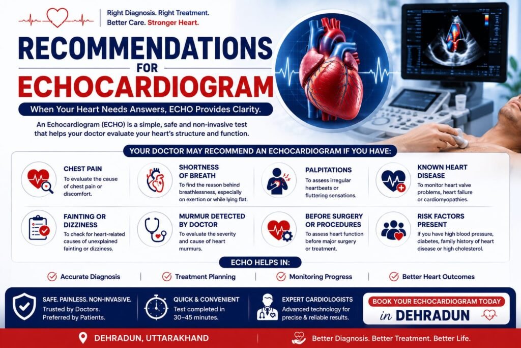
Investigating symptoms such as breathlessness, chest pain, palpitations, dizziness, or fainting.
A heart murmur detected during a routine examination, to check whether it’s coming from a valve problem.
Monitoring known heart failure, to track how well the heart is pumping over time and guide medication decisions.
Following up known valve disease, to monitor whether it’s stable or progressing.
After a heart attack, to assess how much heart muscle was affected and how well the heart is recovering.
Before certain surgeries, as part of a general fitness and risk assessment.
As part of a routine heart checkup, particularly for people with risk factors like high blood pressure, diabetes, or a family history of heart disease, even without specific symptoms.
Monitoring certain medications, in select cases, such as some chemotherapy drugs that can affect heart function over time.
How the Test Is Done
No special preparation needed — unlike some other cardiac tests, a standard echo doesn’t require fasting or any specific preparation.
Lying down — you’ll lie on your left side on an examination table, which brings the heart closer to the chest wall for clearer images.
Gel and probe — a small amount of gel is applied to the chest, and a handheld probe (transducer) is moved across different areas to capture images from multiple angles.
Duration — the test itself usually takes about 20 to 40 minutes, depending on how many views and measurements are needed.
No discomfort — there’s no pain involved; you may feel slight pressure from the probe, but nothing more.
Immediate results — a cardiologist can often review the key findings with you shortly after the test, sometimes the same day.
There’s no recovery time needed — most patients can go straight back to their normal activities immediately after the test.
Understanding Your Echo Report
Echo reports can look intimidating with their list of numbers and abbreviations, but a few terms come up most often:
Ejection fraction (EF) — usually expressed as a percentage; a normal EF is generally in the range of 55–70%. A lower number suggests the heart isn’t pumping as strongly as it should.
LVH (left ventricular hypertrophy) — thickening of the heart’s main pumping chamber wall, often related to long-standing high blood pressure.
Valve gradients — measurements that indicate how much a valve is narrowed, if at all.
Regurgitation grading — usually described as mild, moderate, or severe, indicating how much a valve is leaking.
Diastolic function — describes how well the heart relaxes and fills with blood between beats, separate from how strongly it pumps.
It’s worth being clear that an echo report is meant to be interpreted alongside your symptoms and overall clinical picture by your cardiologist — the same finding can mean different things for different patients, so it’s not something to try to fully self-diagnose from the report alone.
Echo Test Cost in Dehradun
A standard 2D echocardiogram is one of the more affordable cardiac tests, generally costing between ₹1,500 and ₹3,500 depending on the facility and whether additional measurements like Doppler studies are included. This is significantly less than tests requiring a catheter, like angiography, since echo is entirely non-invasive. Exact pricing can be confirmed at the time of booking your test.
Frequently Asked Questions
What is an echocardiogram, and what does it check?
It’s an ultrasound scan of the heart that shows its structure and how well it’s functioning in real time — checking pumping strength, valve function, chamber size, and overall heart muscle movement.
Is an echocardiogram the same as an ECG?
No — an ECG records the heart’s electrical activity as a line graph, while an echocardiogram uses ultrasound to create actual moving images of the heart. They check different things and are often done together for a fuller picture.
Is an echocardiogram painful or does it involve radiation?
No to both. It’s a painless ultrasound test using sound waves, with no radiation involved, and no needles or injections needed for a standard echo.
How long does an echocardiogram take?
Usually about 20 to 40 minutes, depending on how many views and measurements are needed for your specific case.
Do I need to prepare for an echocardiogram?
No special preparation is needed — no fasting, and you can take your usual medications before the test.
What is ejection fraction, and what is a normal range?
Ejection fraction is the percentage of blood pumped out of the heart’s main chamber with each beat. A normal range is generally 55–70%; a lower number can indicate reduced pumping function, which your cardiologist will interpret alongside your symptoms.
How much does an echocardiogram cost in Dehradun?
A standard 2D echo typically costs between ₹1,500 and ₹3,500, depending on the facility and the specific measurements included.
Can an echocardiogram detect a heart attack?
It can show areas of the heart muscle that aren’t moving normally, which often indicates damage from a past heart attack, though the diagnosis of an active heart attack usually relies on ECG changes and blood tests as well.
Can an echocardiogram detect valve problems?
Yes — it’s the primary test used to assess heart valves, showing whether a valve is narrowed, leaking, or functioning normally, and how severe any problem is.
How often should I get an echocardiogram done?
This depends entirely on your individual situation — someone with no symptoms or risk factors may never need one, while someone with known valve disease or heart failure may need one every six to twelve months. Your cardiologist will advise a schedule based on your specific case.
Is a 3D echo different from a 2D echo, and do I need it?
A 3D echo provides more detailed spatial images, useful for certain complex valve assessments or before some procedures. Most routine evaluations are well served by a standard 2D echo, and your cardiologist will advise if a 3D study is needed for your specific situation.
Can an echocardiogram be done during pregnancy?
Yes — since it uses ultrasound and no radiation, it’s considered safe during pregnancy and is sometimes used to monitor heart function in expectant mothers with existing heart conditions.
Whether your doctor has recommended an echo to investigate a symptom, or you’d simply like a clear picture of your heart’s structure and function as part of a general checkup, Your Cardiologist performs and interprets echocardiograms for patients in Dehradun, alongside guidance on what the findings mean for your specific health.